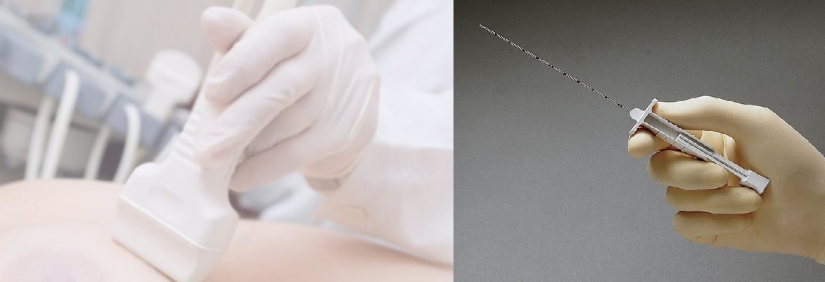
Biopsy is the removal of tissue from any part of the body so that it can be examined for the determination of the existence of a disease. Sometimes a small tissue is taken with the help of a needle, and sometimes suspicious nodule or lump is removed through a surgical operation. The biopsy procedure will depend on the location of the tissue that should be examined. Biopsies can be implemented with imaging guidance such as ultrasound, X-ray, computed tomography (CT) and magnetic resonance imaging (MRI). These types of imaging are used to determine exactly where to place the needle and perform the biopsy.
Biopsy can be applied on every organ like lungs, kidneys, liver, prostate, breast or thyroid. The logic is the same for almost all of the biopsies. After local anesthesia, the needle is moved inside the lump and a tissue is removed. Breast, thyroid or prostate biopsies can be cited as examples.
Some of the main biopsy types are as follows:
- Thyroid biopsy
- Prostate biopsy
- Breast lesion marking
- Breast biopsy
- Thorax biopsy
- Percutaneous nephrostomy
- Abscess drainage
- Abdominal Organ Biopsies
Thyroid biopsy

Thyroid biopsy is completed within 10-15 minutes with ultrasound guidance. Thanks to the usage of US to guide the process, the risk of damaging the veins or other organs is eliminated. A pathology expert also attends the process and carries out microscopic evaluation. Then the expert informs the radiology expert about whether or not the tissue taken is enough to make a decision. This way, a repeat of the process will not be needed and the action will be completed in a safe and reliable manner.
Prostate biopsy
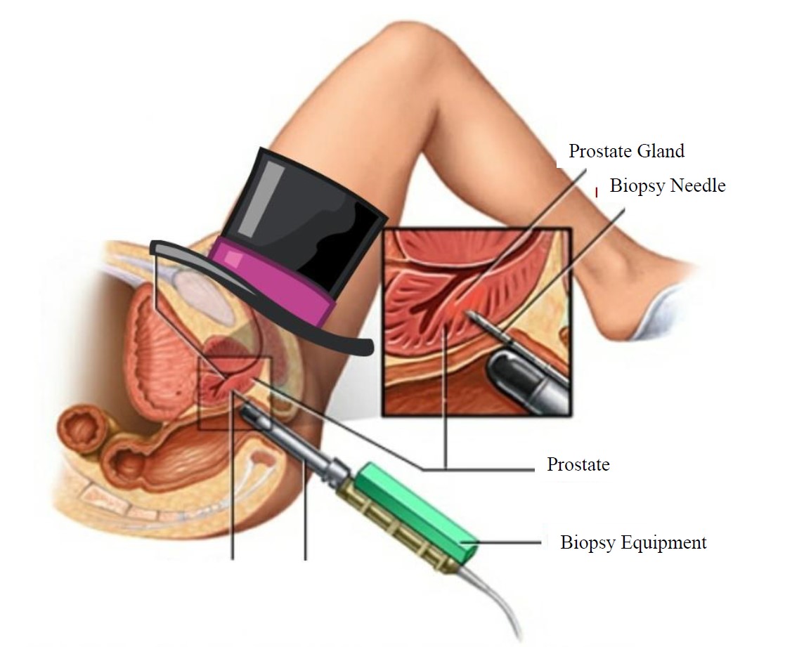
This is done to remove tissue from the prostate with ultrasonography guidance so that a prostate cancer can be diagnosed. After histopathological examination is done on the tissue taken from the prostate, a pathology expert will announce the prostate cancer diagnosis according to the results. The procedure is done through rectum. A special probe will be inserted inside the rectum and the prostate will be examined through ultrasonography. Procedure is done under local anesthetics. After ultrasonographic evaluation, a minimum of six, 1 mm diameter, and 15-20 mm long tissue samples will be taken from pre-determined areas. There will be no pain during the procedure due to local anesthetics.
Fine-Needle Breast Biopsy

For abnormal breast formations which cannot be felt by hand (and which can be detected only through mammography or US), guide wire marking is applied in addition to FNBB, core and vacuum-assisted core biopsy.
Fine-needle aspiration biopsy takes around 15 minutes, is very easy and painless. It doesn’t require preparation. The biopsy materials will be sent to the pathology laboratory and pathology results will be obtained within 4-5 days. Fine needle biopsy has no risk when done by experienced people, and no complications are expected. The process will be ended in a safe and reliable manner.
Thorax Biopsy
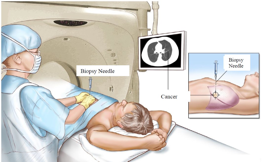
In this invasive procedure, a biopsy needle is placed inside the tissue to take tissue samples from the chest wall or from the lungs and is done with the help of CT. It is important that you don’t eat within the last six hours before the procedure. However, you are free to take your medications. If you have diabetes, please consult your physician for the dosage of your insulin.
You should go to the examination area 1 hour before the procedure so that the preparations can be made. The stuff in the radiology department may ask for some blood tests. They will also give you details about the procedure and may ask you to sign a consent paper.
The procedures might take from 30 minutes to 90 minutes depending on the size of the relevant area and the number of the tissue samples to be taken. You might be kept under supervision for 2-4 hours to ensure that no complications arise.
Abdominal Organ Biopsies
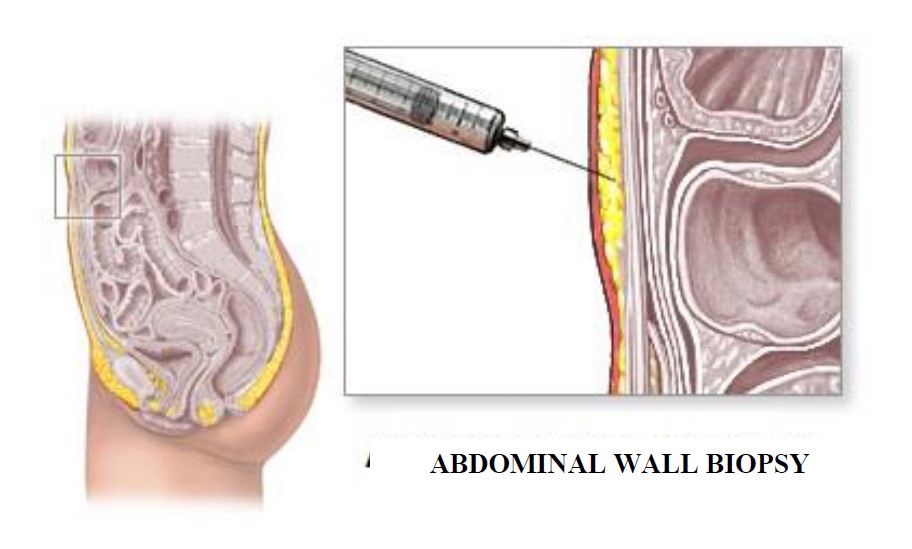
Biopsy can be applied on almost every organ in the abdominal area. Abdominal biopsy is used to determine whether a lump in the abdominal area is cancerous or benign. The lumps can be located in the fat, deep within the abdomen. A sample of the lump is removed percutaneously under image guidance (ultrasound or CT), or surgically using a laparoscope or by open surgery.
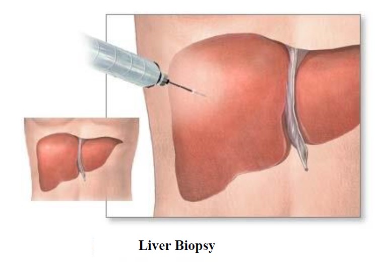
Liver biopsy is used for the diagnosis of various diseases like hepatitis C, cirrhosis, infections and cancer in the liver. Also it can be used to determine if an implanted organ will be rejected by the body or not. Usually, a needle will be percutaneously inserted through the skin. Biopsy can also be done through a catheter placed through jugular vein or can be done through a surgical procedure.
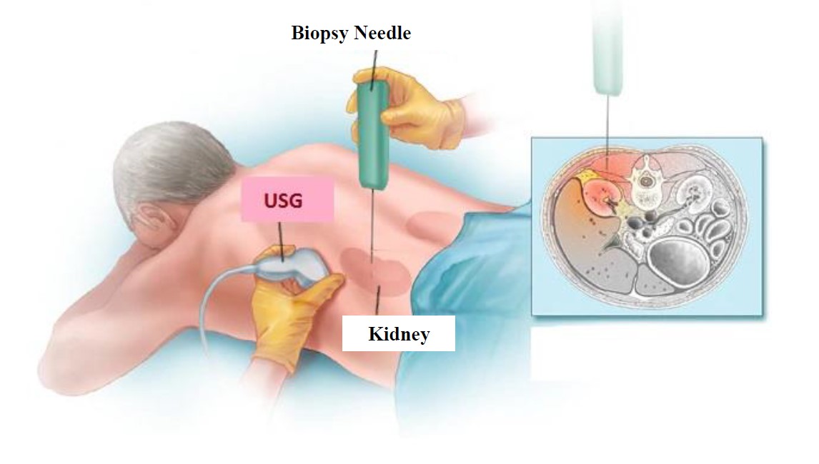
Kidney biopsy, is used to determine kidney failure, kidney infections or to examine is suspicious tumor (like cancer). It can also be used to determine if a newly implanted transplanted kidney will be rejected or not. Kidney biopsies will be done with imaging guidance (ultrasonography or CT) in order to remove a small tissue sample.
Pancreas biopsy can be done with ultrasonography or tomography guidance. The imaging technology that shows the lump the best should be chosen. A guide needle will be used to reach the lump in the pancreas and then a fine biopsy needle will be used to take multiple samples. It is important that the guide needle does not contact the veins, bowels, and abdomen and the small intestines. For this reason, it is important to use tomography or ultrasound guidance. When done by experienced people, the procedure is painless, safe and takes only 10 minutes.
Colon biopsy is done to remove and examine colon tissues. It is intended to determine whether a tissue is cancerous or pre-cancerous.
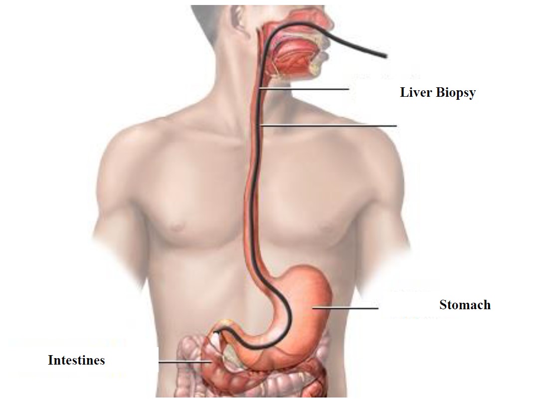
Standard biopsy method for abdomen, small intestines and colon is the endoscopic biopsy. It is done by gastroenterology or general surgery, and the procedure will be done through the mouth, or rectum and multiple samples will be taken from the abdomen, colon or the intestines. This biopsy method is suitable for the masses that grow towards the lumen. But sometimes abdomen-intestine masses grow out and since they cannot be seen with endoscopy, they cannot be cut out with conventional methods. In such cases, percutaneous needle biopsy can be applied under tomography guidance for a diagnosis.
Percutaneous nephrostomy

A percutaneous nephrostomy is the placement of a catheter through your skin into your kidney to drain your urine. It is inserted through your back or flank. This is done to reduce to pressure on the kidneys when there is a problem that completely or partially prevents the flow of urine or when there is urine leakage into the abdomen.
Abscess drainage
Abscess is the collection of pus or infection in any part of the body. These abscesses are reliably drained without surgery, with imaging guidance using catheter.