
Renal cancer is not a common type of cancer, however, its incidence has increased in recent years; it is important because it is resistant to chemotherapy, radiotherapy and hormonal therapy. Today, the main treatment in kidney cancers is surgical procedures, removing all or a part of the kidney and surrounding tissues and targeted smart drugs. Smart drugs and immunotherapies are effective in advanced (4Th) stage renal cancers. However, these procedures could not always be applied in several conditions such as underlying diseases, limited renal functions, having a single kidney, the metastasize of the cancer outside the kidney, and older age. Therefore, alternative treatments are being studied.
Thanks to the developments in today's technology, the number of alternative treatment methods for small renal tumors is increasing. Therefore, several methods were developed for renal cancer treatments. Small renal cancers may sometimes be destroyed by non-surgical treatments such as hot and cold. These procedures may be a very good option for people with health problems posing a risk for surgery.
Non-surgical treatment options may include:
- Treatment by freezing cancer cells: cryoablation therapy. During cryoablation, a hollow needle specific to renal tumor is inserted through your skin by ultrasonography or other imaging guides. The cold gas in the needle is used to freeze cancer cells.
- Treatment by warming cancer cells: radiofrequency ablation therapy (RFA). During radiofrequency ablation, a probe specific to renal tumor is inserted through your skin by ultrasonography or other imaging guides. An electrical current is applied from the needle to the cancer cells, causing the cells to heat or burn.
Radiofrequency Ablation Therapy (RFA)

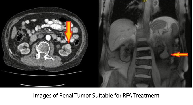
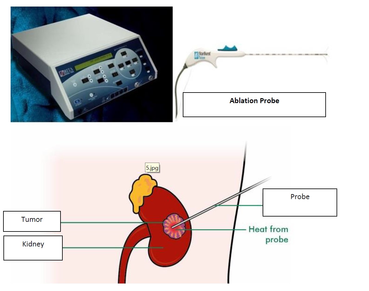
Radiofrequency ablation therapy is a treatment method that uses high-energy radio waves to warm the tumor. A thin, needle-like probe is inserted into the skin and moved forward till its tip is located into the tumor. The insertion of the probe is guided by ultrasonography or CT (Tomography) scanning. Once inserted, an electric current is applied through the probe tip. This warms the tumor and destroys the cancer cells.
The aim of RFA treatment is to cause cell death and coagulation necrosis in tumor tissue by heating the tumor tissue to a very high tissue temperature. RFA is a hyperthermic ablation therapy method approved by FDA (US Food and Drug Administration) for the treatment of soft tissue tumors. It may also be applied successfully in liver, kidney, lung, bone and breast tumors.
RFA treatment may be applied both laparoscopically and percutaneously, so the patients may be minimally treated invasively.
In the laparoscopic transperitoneal method the tumor tissue is removed after the colon is dissected. Tumor size is determined by laparoscopic ultrasonography probe. Before RFA, biopsy should be performed by an 18 G Tru-Cut needle. The RFA probe is inserted perpendicular to the tumor surface and its teeth are opened. After rechecking the teeth of the RFA probe by ultrasonography, ablation was performed. An ablation zone is formed up to 0.5 cm to tumor border.
Percutaneous approach may be applied under sedation or general anesthesia, guided by ultrasonography or computed tomography. RFA treatment under general anesthesia guided by computed tomography is safer. Tumor size and extension are determined. Before RFA, tumor biopsy should be performed by an 18 G Tru-Cut needle. The RFA probe is inserted in the tumor tissue and an ablation zone is formed up to 0.5 cm to the tumor tissue.
Which common uses of the procedure are preferred?
Radiofrequency ablation is primarily used to treat renal cell carcinoma (kidney tumors).
Ablation is a feasible and effective treatment option in the following conditions:
- For cancer treatment in individuals who continue life with a single kidney,
- In patients with other medical conditions contraindicated for surgery,
- In elderly age and patients who may have difficulty in recovery after surgery
- In patients with tumors smaller than 4 centimeters,
- With tumors in both kidneys or a familial predisposition (family history) to more than one renal tumors,
- Patients with a recurrent tumor after surgical resection,
Ablation therapy is preferred. It may be also used as a preoperative method to reduce blood loss during surgery.
How to Perform
Firstly, you will be asked not to eat or drink anything after midnight before the procedure. Your doctor will tell you the medications to be taken in the morning. Your blood may also be tested to determine the functional condition of your kidneys and your clotting status.
The procedure is usually performed by the guidance of an imaging method (USG or CT) in an interventional radiology room or sometimes in the operating room by a specialist radiologist. Women should always inform their doctor and x-ray technician if there is a risk of pregnancy. Several imaging tests are contraindicated during pregnancy to avoid radiation exposure to the fetus. If x-rays are required, several precautions will be taken to minimize radiation exposure to the baby.
Ablation is usually performed as an outpatient treatment protocol. You will be asked to wear an apron and to lie on the procedure table. You may be connected to monitors to follow up your heart rate, blood pressure, oxygen level, and heart rate. A nurse or technician will insert an intravenous line into a vessel in your hand or arm to administer intravenous (IV) sedation medication. In several cases, the anesthesiologist may manage your sedation or apply general anesthesia.
Several tumors are located close to the urine collecting area of the kidney or in the ureter. In such cases, your urologist may insert a temporary stent from your urethra to the ureter. During ablation, cold water is slowly dripped from this stent to protect these structures against heat damage. The stent is generally removed at the end of the procedure.
The needle insertion area will be sterilized and closed with a sterile cover. Your doctor will administer local anesthesia into the area. This procedure may burn or irritate the area immediately before the area becomes anaesthetized. A very small skin incision is made in the area. If more than one needle is needed, multiple notches may be made.
Guided by imaging guide, your doctor will insert the needle electrode through the skin and move forward to the tumor area. Energy is applied after the needle electrode is inserted. For a large tumor, multiple ablation may be needed by repositioning the needle electrode or inserting multiple needles in different parts of the tumor to prevent remaining tumor tissue behind.
At the end of the procedure, the needle electrode is removed and pressure is applied to stop any bleeding, and a dressing is applied to close the opening in the skin. No sutures are required.
Each ablation lasts approximately 10 to 30 minutes, and an additional time is required for more than one ablation. The whole process is generally lasted 1 to 3 hours.
After RFA treatment, the patients should be screened by contrasted computed tomography at 6th week and 6th month, chest graph, alkaline phosphatase measurement, liver function tests, physical examination twice a year. In magnetic resonance imaging or computed tomography in the first sixth week after RFA, an increase in the ablation zone is defined as incomplete ablation. If there is a recurrence in the ablation zone or incomplete ablation, re-ablation or other surgical treatments may be applied.
Several advantages of the RFA method
- Ablation is a relatively fast procedure and recovery is fast, thus may be continued almost immediately in patients who need chemotherapy.
- Ablation is cheaper than other treatment options.
- A surgical incision is not required - a small notch in the skin without the requirement of suture is enough.
- Unlike surgical treatment, this procedure protects kidney.
- It has no effect on blood pressure.
- Depending on the width of the area to be treated, it may have an effect on kidney function or not.
In conclusion, in parallel with the advanced technology, RFA treatment has been introduced as an alternative treatment method in the treatment of patients with small kidney tumors and is becoming increasingly common.
RFA treatment may be applied both laparoscopically and percutaneously, allowing the patients to be treated minimally invasive. Short recovery time, decreased morbidity rate and reliable oncological findings in the long-term follow-up period are very promising findings for the common use of RFA treatment.
Cryoablation Method

It is observed that cryoablation (freezing to destroy) treatment has a very high effect in certain groups of with renal cancer.
However, these procedures could not always be applied in several conditions such as underlying diseases, limited renal functions, having a single kidney, the metastasize of the cancer outside the kidney, and older age. Therefore, alternative treatments are being studied. Among them, cryoablation is a method that has introduced in recent years with successful results. In this method, the tumor is rapidly cooled and frozen to be destroyed with the help of needles inserted through a small hole in the skin.
The 5 year renal cancer specific survival rate results of several studies assessing the efficacy of cryoablation in renal cancer were found to be similar in cryoablation and partial nephrectomy surgery, an operation to excise some part of the kidney. When the side effects were compared between the procedures; cryoablation was more advantageous in terms of non-urological side effects in 30 days. Besides, the incidence of renal failure was found to be lower in cryoablation as compared to partial nephrectomy.
In conclusion, it was observed that cryoablation technique had a very high efficacy and safety profile in the treatment of certain patient groups with renal cancer, deserving to be much more involved in clinical practice. However, this method is not suitable for all renal cancers. In tumors up to 4 cm, its efficacy rate is about 98%, but it is not effective enough in bigger tumors. Therefore, it is very important to determine the patient groups eligible for cryoablation method thoroughly and it should be performed in experienced centers.